
Visible spider veins are not a niche cosmetic complaint. When spider telangiectasias and reticular veins are counted alongside varicose veins, superficial venous changes have been reported in 80% of men and 85% of women, while an independent Scottish sample found spider veins in 88% of women and 79% of men. Most symptoms are mild, but the right treatment still depends on more than appearance. StatPearls summarizes the epidemiologic and clinical context.
Removing spider veins safely means matching the treatment to the vessel's size, depth, location, your skin tone, and any symptoms that suggest venous reflux. Sclerotherapy remains a central option for symptomatic leg veins, while laser and light-based treatments can be useful for very small vessels or as an adjunct. The important question isn't only which treatment is most advertised. It's which problem you're treating.
Table of Contents
- What Spider Veins Are and Why They Form
- Proven Treatment Options to Remove Spider Veins
- Choosing the Right Treatment for Your Vein Type
- What to Expect During and After Your Procedure
- When Spider Veins Signal a Deeper Venous Problem
- Prevention Strategies and Choosing a Qualified Provider
- Your Next Steps Toward Clearer Skin
What Spider Veins Are and Why They Form
Spider veins are visible in many forms: a red fan near the ankle, blue lines behind the knee, or a purple web across the thigh. These small, widened superficial vessels are called telangiectasias. Reticular veins are larger blue-green vessels that may feed them, while varicose veins are raised and more prominent, requiring a different assessment.
The underlying issue is often increased pressure in the superficial venous network. Veins return blood toward the heart with help from valves and surrounding muscle movement. If the valves or vessel walls work poorly, pressure can build and cause vessels near the skin to widen. Genetics, hormonal changes, pregnancy, obesity, prolonged standing, and age-related changes may all contribute. Risk also increases with age, pregnancy, obesity, and work that involves standing for long periods. The clinical overview in StatPearls discusses these risk factors and the widespread nature of visible superficial veins.
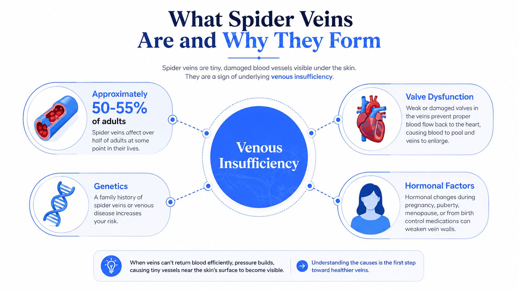
Appearance and symptoms don't always match
Some patients seek treatment solely because they dislike the appearance. Others notice aching, heaviness, itching, or fatigue in the affected leg. The same surface pattern can therefore be a cosmetic concern for one person and a sign of a broader venous problem for another.
Vein diameter helps determine the treatment pathway. Very fine telangiectasias may respond well to light-based treatment, while larger reticular veins often respond more predictably to injection therapy. Skin tone also affects the safety discussion, because pigment changes can be more concerning with certain energy-based treatments. Raised veins, swelling, or persistent heaviness warrant a fuller evaluation before treating the visible surface vessels.
Clinical rule: Clearer skin is only one treatment goal. A sound consultation also checks whether pressure from a deeper vein is contributing to the visible network.
The cause and pattern matter. Treating every visible line with the same modality can leave feeding veins untreated or expose the skin to an unsuitable approach. A sound plan considers the vessel's diameter, depth, location, skin tone, symptoms, and the condition of the surrounding venous system.
Proven Treatment Options to Remove Spider Veins
Sclerotherapy is generally the first procedural option for symptomatic spider veins and reticular veins. A clinician injects a liquid or foam sclerosant into the vessel. The solution injures the vessel lining, prompting fibrosis and eventual closure. For spider veins, commonly used concentrations include 0.1% to 0.2% sodium tetradecyl sulfate or 0.25% to 0.5% polidocanol, as outlined in the intersocietal sclerotherapy protocol. The appropriate concentration still depends on vessel size, location, and the clinician's technique.
Laser treatment sends focused energy through the skin to heat and collapse selected vessels. It can suit very small, superficial telangiectasias, either as an alternative to injections or alongside them. Evidence reviewed by Cochrane found similar improvement or resolution between laser and sclerosing agents in relevant studies, while some comparisons showed less hyperpigmentation after laser. The outcome depends on wavelength, cooling, energy settings, vessel depth, and skin pigmentation. The Cochrane review discusses laser treatment, sclerotherapy, and pigmentation trade-offs.
Light-based treatment can address fine, superficial vascular redness. It is not interchangeable with laser or sclerotherapy, because its usefulness depends on how superficial the vessels are and how the patient's skin responds to energy. Broadband Light therapy information may help explain this category, but an examination must determine whether visible vessels are suitable.
Matching the modality to the vessel
| Treatment | How it works | Best fit | Main limitation |
|---|---|---|---|
| Sclerotherapy | Injected sclerosant damages the vessel lining and closes the channel | Small spider veins and reticular veins | Temporary swelling, bruising, or pigment change may occur |
| Laser therapy | Focused light heats and collapses the vessel | Very fine telangiectasias | Requires careful settings, particularly with darker skin tones |
| Light-based therapy | Broad or targeted light treats superficial vascular redness | Fine, superficial vessels | Results vary with vessel depth, skin type, and treatment settings |
Very fine vessels may respond better to laser or light-based treatment. Larger reticular veins usually require injection therapy, especially when they feed a visible network. A clinician should also assess whether a prominent branch is part of a deeper varicose pattern before treating it as an isolated cosmetic vessel.
Repeated sessions may be needed with any modality. Clearance depends on vessel diameter, depth, distribution, skin tone, and the treatment response, so a fixed clearance rate would be misleading. Pigment risk deserves a direct discussion before energy-based treatment, particularly for darker skin tones.
Topical vitamin K, arnica, apple cider vinegar, dry brushing, and retinoids are not substitutes for procedural treatment. They may have limited supportive uses, but they do not close an abnormal vein like a properly delivered sclerosant or vascular laser. Combination treatment can make sense when very fine surface vessels coexist with larger reticular feeders. Practice teams can review administrative considerations in the Happy Billing dermatology page, while patients should base treatment decisions on diagnosis, vessel characteristics, and safety rather than billing terminology.
Choosing the Right Treatment for Your Vein Type
A vein under 1 mm may respond well to laser, while larger vessels or those connected to a reticular feeder often call for injection therapy. The first decision is therefore the vessel itself, not the device. A visible branch may also belong to a deeper varicose network, so it should not be treated as an isolated cosmetic telangiectasia without assessment.
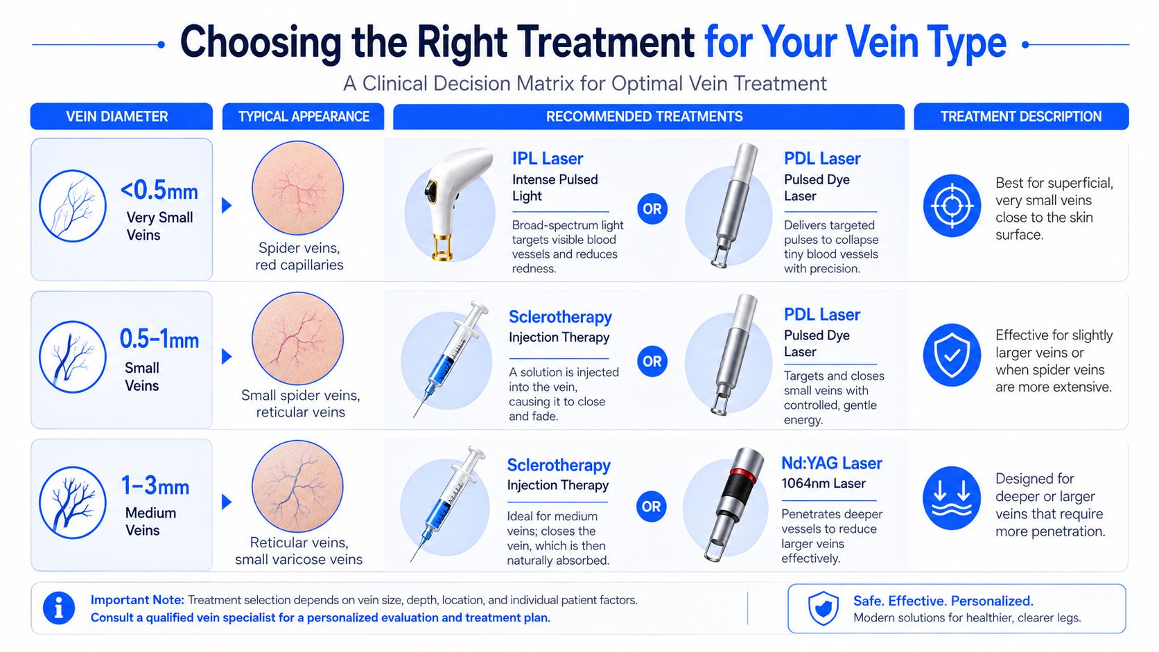
The practical decision points
- Very fine vessels: Laser or light-based treatment may suit tiny, superficial vessels. Skin tone, vessel depth, and the risk of post-inflammatory pigmentation still determine whether the expected benefit justifies the treatment.
- Small to moderate leg veins: Sclerotherapy is often the more direct option because the solution enters the vessel lumen. It becomes more relevant when aching, heaviness, or an underlying feeder is also present.
- Darker skin tones: Wavelength, cooling, and energy settings require careful planning because pigmentary changes can follow energy-based treatment. ProMD's guide to laser treatment for darker skin provides general background, but a trained clinician must select settings for the individual patient.
- Facial vessels: Surface laser or light-based treatment is generally considered more suitable than injecting sclerotherapy into facial vessels, where anatomy and injection risk require particular caution.
- Symptoms or recurrence: Swelling, persistent aching, heaviness, or rapid recurrence changes the consultation from a cosmetic discussion to a venous assessment.
A patient with isolated, fine, symptom-free vessels may reasonably prioritize minimal downtime. A patient with symptomatic leg veins may accept compression and staged treatment because surface clearance does not address the wider clinical picture. One modality should not be selected automatically for both.
Treatment planning also depends on distribution. Scattered fine vessels may need a different approach from a dense network supplied by larger reticular veins. Combination treatment can be appropriate when both patterns are present, provided the clinician identifies the feeder and sets realistic expectations about repeat sessions.
Ask this before booking: “What makes this modality appropriate for my vessel size and skin tone, and what would make you choose something else?”
One-sided swelling, significant pain, skin changes, a history suggestive of clotting, or prominent varicose veins warrant medical evaluation before an aesthetic treatment plan is finalized.
What to Expect During and After Your Procedure
A thorough appointment begins with an examination and treatment mapping. The clinician cleanses the skin, identifies the visible vessels, and decides whether injections or a light-based handpiece is more appropriate. Medication changes, sun exposure, and skincare products should be reviewed in advance rather than managed casually on the day of treatment.
For sclerotherapy, the patient lies horizontally while the provider uses a fine needle to place a small amount of sclerosant inside the target vessel. Fine 30 to 32 gauge needles and low sclerosant concentrations help limit extravasation and tissue injury. For laser treatment, the handpiece delivers controlled light or laser energy over the vessel, often producing a mild snapping or stinging sensation. The procedural review describes the mechanism, technical approach, horizontal positioning, and compression considerations.
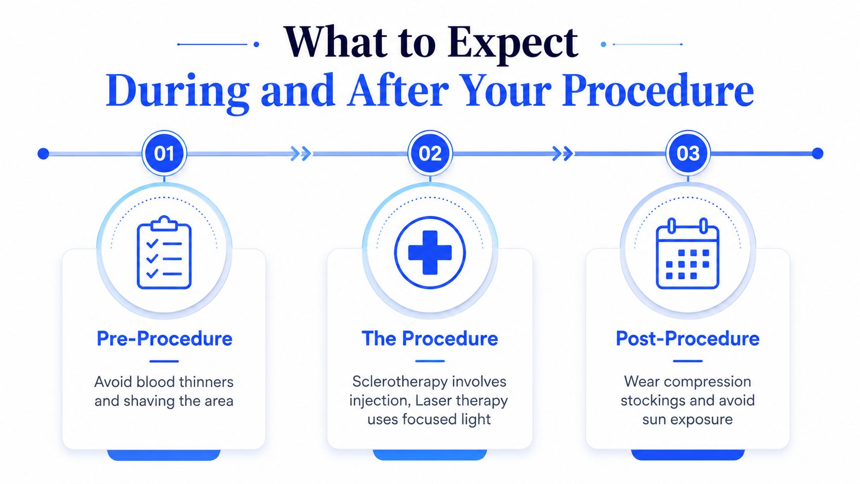
Aftercare affects the result
Compression is not an optional detail after many leg-vein treatments. Guidance describes compression around 23 to 32 mm Hg for up to three weeks after sclerotherapy, and the German S2k guideline states that compression after liquid sclerotherapy improves clinical response and reduces hyperpigmentation. The German guideline explains the role of horizontal positioning, low-friction syringes, and post-treatment compression.
Patients are commonly encouraged to walk after treatment because gentle movement supports circulation. They should follow their provider's specific instructions about strenuous exercise, hot baths, saunas, and sun exposure. Bruising, temporary redness, trapped blood, superficial phlebitis, and hyperpigmentation can occur. Pigment changes may take weeks or longer to fade, especially when the skin is more prone to post-inflammatory pigmentation.
A treatment series is often planned rather than promised as a single visit. The interval depends on how the vessels respond and whether residual feeders remain. The guide to Halo laser recovery provides general recovery-planning context for laser-treated skin, but spider-vein aftercare must follow the actual modality used.
Contact the treating provider for worsening pain, marked swelling, spreading redness, significant skin injury, or symptoms that feel out of proportion to routine bruising and irritation.
When Spider Veins Signal a Deeper Venous Problem
Spider veins aren't automatically evidence of a serious vascular disorder, and asymptomatic telangiectasias don't routinely require duplex ultrasound. The clinical threshold changes when symptoms are present. The 2023 Society for Vascular Surgery and American Venous Forum guideline recommends evaluating symptomatic chronic venous disease for reflux, while symptomatic telangiectasias and reticular veins are treated with liquid or foam sclerotherapy. The guideline summary outlines when ultrasound is and isn't routinely needed.
Symptoms that change the plan
Seek a vascular assessment before cosmetic treatment if you have:
- Persistent heaviness or fatigue: Symptoms that build during the day may indicate more than an isolated surface concern.
- Swelling or aching: Especially when symptoms worsen after standing.
- Skin changes: Discoloration, dermatitis, thickening, or other changes around the lower leg deserve evaluation.
- Clusters or recurrence: Repeated return in the same area can suggest an untreated source or feeder.
- Larger veins: Raised, winding varicosities require a different assessment from fine telangiectasias.
A venous reflux study uses duplex ultrasound to assess blood flow and identify backward flow through incompetent valves. If reflux is present, the clinician may need to address the source before treating the surface network. Treating only the visible vessels can produce disappointing results when deeper pressure continues to feed them.
Escalate the consultation when the aesthetic concern comes with swelling, heaviness, aching, skin changes, or recurrent vessels. A surface procedure shouldn't substitute for a vascular workup when symptoms point deeper.
The distinction protects both your health and your investment in treatment. A patient with asymptomatic spider veins may proceed through an aesthetic pathway, while a patient with symptomatic chronic venous disease may need vascular medicine or vascular surgery input first.
Prevention Strategies and Choosing a Qualified Provider
Prevention can reduce strain on the venous system, but it can't erase inherited susceptibility or guarantee that new vessels won't form. Regular walking and calf-muscle activation help support venous return. Ankle flexion during periods of sitting, changing position during long standing periods, maintaining a healthy weight, and using graduated compression when clinically appropriate are practical measures.
Compression is most useful when it fits the patient's circulation and symptoms. A clinician can advise whether graduated hosiery makes sense, especially for people who stand for work, experience leg fatigue, or have a history of venous problems. Topical creams and supplements should be treated cautiously. Evidence for cosmetic products such as vitamin K creams or anecdotal remedies is limited compared with procedural evidence, and supplements may interact with medications or medical conditions.
What qualified care looks like
Look for a provider with relevant board certification and hands-on experience in dermatology, vascular medicine, vascular surgery, phlebology, sclerotherapy, or vascular laser treatment. The practice should be able to explain:
- Why the modality fits: The recommendation should reflect vessel diameter, location, symptoms, and skin tone.
- How complications are handled: Ask about pigmentation, matting, trapped blood, phlebitis, and skin injury.
- Whether ultrasound is available: Symptomatic patients may need reflux evaluation before surface treatment.
- Who performs the procedure: Confirm the treating professional's credentials and physician oversight.
A discount-driven setting may emphasize a device or package without adequately addressing the source of recurrent veins. You should leave the consultation understanding what the provider plans to treat and what the plan won't address.
ProMD Connect can provide a convenient virtual starting point for an initial assessment. A teleconsult may help determine whether a straightforward cosmetic concern appears suitable for an in-clinic evaluation, whether symptoms suggest the need for in-person assessment, and how treatment scheduling could proceed. Because spider veins can require direct examination or duplex ultrasound, virtual care should guide the next step rather than replace testing when reflux is suspected.
Your Next Steps Toward Clearer Skin
A successful plan to remove spider veins depends on vein diameter, skin phototype, location, symptoms, and underlying venous health. The most visible treatment isn't automatically the safest or most effective choice for your anatomy.
Start with a simple self-assessment. Note whether the vessels are fine or raised, red or blue, isolated or clustered, and whether you have heaviness, aching, itching, swelling, or skin changes. Next, choose the appropriate entry point. A teleconsult may be reasonable for a straightforward cosmetic concern, while symptoms or recurrence warrant an in-person evaluation and possible duplex ultrasound.
Bring focused questions to the consultation:
- Which modality fits my vessel size and skin tone?
- Are there feeder or refluxing veins that should be evaluated?
- What treatment sequence do you recommend?
- What compression protocol will I need afterward?
- How will you manage pigmentation, bruising, matting, or trapped blood?
Sclerotherapy and laser can both be effective, but neither removes the possibility of new veins developing elsewhere. Prioritize a board-certified dermatologic or vascular clinician who explains trade-offs clearly instead of choosing a provider based only on a discounted package.
ProMD Health offers non-surgical aesthetic services, including laser and Broadband Light therapy, with consultation pathways designed to help determine whether a visible vascular concern is appropriate for in-clinic treatment or needs further evaluation. Visit ProMD Health to explore available services and request the next step for your spider-vein concerns.


