
The most popular advice for hyperpigmentation around the mouth is often the least useful: apply a brightening cream and wait for the area to fade. That approach assumes every dark patch is melasma or ordinary excess melanin. In practice, the same perioral zone can show post-inflammatory hyperpigmentation, contact dermatitis, constitutional pigmentation, vascular discoloration, or a structural shadow caused by facial volume and anatomy.
That distinction matters because the wrong treatment can prolong irritation and make pigment more noticeable. A clinician first asks what changed, what has been touching the skin, whether the area has been inflamed, and whether the darkness is pigment rather than shadow. The treatment should follow that diagnosis, not the other way around.
Table of Contents
- Why Perioral Darkening Is Rarely Just Melasma
- Identifying the Root Triggers of Perioral Pigmentation
- Evaluating Pigment Depth and Skin Phototype
- Evidence-Based Clinical Treatments for Dark Spots
- Building a Preventative Medical-Grade Skincare Routine
- When to Seek Professional Dermatologic Evaluation
Why Perioral Darkening Is Rarely Just Melasma
Melasma can extend onto the upper lip and around the mouth, but it shouldn't be the automatic diagnosis. Perioral hyperpigmentation is a location, not a disease, and several different conditions can occupy that location at the same time. The differential includes melasma, post-inflammatory hyperpigmentation from acne or eczema, pigmented contact dermatitis, drug-induced pigmentation, constitutional pigmentation, smoking-related changes, and, rarely, a lesion that requires biopsy rather than cosmetic treatment. A clinical discussion of pigmentation around the mouth supports treating the phrase “around mouth” as a prompt for differential diagnosis.
Melasma often has a broad, relatively even distribution, while post-inflammatory pigment tends to follow the exact area of a previous eruption, rash, burn, or procedure. Irritation-related discoloration may sit beside redness, scaling, itching, or small bumps. A shadow from volume loss or facial anatomy can look brown in some lighting but won't behave like melanin when viewed closely or treated with a pigment inhibitor.
The danger of a universal brightening routine
A routine designed for melasma may contain multiple pigment-targeting actives. If the actual problem is perioral dermatitis, lip-licking dermatitis, or a reaction to toothpaste or cosmetics, adding more active products can increase barrier disruption. More inflammation creates more opportunity for melanocytes to become stimulated, especially in skin that is already prone to persistent post-inflammatory pigment.
Post-inflammatory hyperpigmentation is a foundational cause of darkening in this region. It follows inflammation or injury from conditions such as acne and eczema, and it can remain after the original problem has settled. The process is straightforward: inflammation signals melanocytes, excess pigment accumulates in the epidermis or dermis, and the visible mark lasts longer than the trigger that caused it. The dermatology review on post-inflammatory hyperpigmentation describes this inflammation-to-pigment pathway across skin types and highlights its greater frequency in many skin-of-color populations.
Clinical rule: Before trying to lighten the mark, identify what keeps irritating the skin.
The first useful question isn't “Which cream removes dark spots?” It's “Is this true pigment, pigment left after inflammation, or a shadow that needs a different approach?” That answer determines whether the priority is trigger removal, topical pigment control, a superficial peel, laser assessment, camouflage, or evaluation of volume and vascular factors.
Identifying the Root Triggers of Perioral Pigmentation
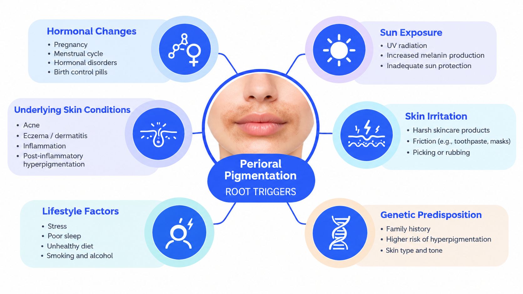
The mouth area is exposed to repeated movement, moisture, friction, product contact, and accidental neglect during sun protection. Those exposures can create low-grade inflammation that isn't dramatic enough to feel like a rash but is sufficient to sustain discoloration. The most productive way to investigate hyperpigmentation around the mouth is to separate inflammatory triggers from contact reactions and structural causes.
Inflammation leaves pigment behind
Acneiform eruptions around the chin and mouth can leave flat brown marks after the bump disappears. Eczema, seborrheic dermatitis, perioral dermatitis, chapping, and lip-licking can create the same outcome. Repeated rubbing, picking, wiping after meals, or applying irritating products keeps the inflammatory signal active.
Hair removal deserves a careful review. Waxing, threading, and poorly controlled device settings can injure or inflame the skin, particularly across the upper lip. The pigment may follow the exact band of contact, which can make it resemble melasma even though the trigger is mechanical irritation. A primary-care dermatology guideline emphasizes that treating the underlying inflammatory disorder promptly can reduce the development of post-inflammatory pigmentation.
Everyday contact can be the hidden cause
Localized darkening may result from allergic or irritant reactions to toothpaste, lip products, saliva, foods, hair dye, or skincare products. A product doesn't have to burn immediately to be relevant. Repeated exposure can produce subtle redness, dryness, tightness, peeling, or itching before the area becomes visibly darker.
Audit exposures in a practical order:
- Oral care: Consider toothpaste and products that repeatedly contact the skin at the corners of the mouth.
- Lip care: Review balms, glosses, tints, plumping products, and fragranced formulas.
- Saliva and licking: Notice whether the tongue repeatedly wets the surrounding skin, followed by drying and friction.
- Hair removal: Map the discoloration against the area treated by waxing, threading, or another method.
- Active skincare: Pause new acids, scrubs, retinoid-like products, and brighteners applied too close to the mouth if irritation is present.
A product history is more valuable than adding another serum when the area is inflamed. Dermatology guidance on hyperpigmentation around the mouth specifically identifies toothpaste, lip products, saliva, foods, skincare, hair removal, eczema, and seborrheic dermatitis as possible contributors.
Structural darkness isn't melanin
A hollow beside the mouth, reduced facial volume, or a vascular undertone can create a darker appearance without primary pigment excess. Lighting changes, facial movement, and the direction of the shadow can offer clues. If the darkness is strongest beneath an overhang or contour and doesn't look like a flat patch under close examination, pigment creams alone may disappoint.
That doesn't mean structural treatment is automatically appropriate. It means diagnosis comes first. A provider may recommend observation, camouflage, anti-inflammatory care, pigment-directed treatment, or a combined plan based on what creates the appearance.
Evaluating Pigment Depth and Skin Phototype
A clinician evaluates where the pigment sits before choosing a peel, laser, or active ingredient. Epidermal pigment is closer to the surface and may respond more predictably to topical therapy and superficial exfoliation. Dermal pigment sits deeper and can be more resistant, while mixed pigment requires a more conservative and combined strategy.
The assessment begins with the pattern. Symmetry may suggest melasma or constitutional pigmentation, while marks that trace previous acne, rash, or hair removal point toward post-inflammatory change. The provider also examines texture and color. Flat brown discoloration behaves differently from gray-brown depth, redness, scaling, papules, or a shadow created by contour.
Why phototype changes the risk calculation
Post-inflammatory hyperpigmentation can occur at any age and in all skin types, but it is more common and more persistent in Fitzpatrick skin types III through VI. In darker-skinned patients with acne, reported incidence can reach 65%, and PIH accounts for up to 8.5% of dermatology consultations, according to the major review indexed by PubMed. Those figures explain why a treatment that seems mild on paper can produce a difficult rebound response in skin of color.
The provider therefore considers baseline pigmentation, previous reactions, current inflammation, recent tanning or sun exposure, and the patient's history with peels or lasers. Device settings and treatment depth must be selected for the individual, not copied from a generic protocol. Conservative sequencing is often safer than pursuing immediate intensity.
A practical diagnostic checklist
Before treatment, ask:
- Is the surface smooth? Smooth, flat pigment suggests a different process from an active rash or textured eruption.
- Does the pattern match a trigger? Previous acne, dermatitis, licking, wiping, or hair removal can reveal the cause.
- Does the color change with lighting? A changing shadow may indicate anatomy or volume rather than melanin.
- Is the area still irritated? Persistent inflammation should be controlled before aggressive pigment treatment.
- Could a medication or product be involved? Drug-induced and contact pigmentation require exposure review, not stronger lighteners.
For readers considering a device-based option, ProMD Health's guide to laser treatment for darker skin provides relevant context on why skin tone and treatment planning must be considered together. A proper consultation can also identify when laser is unlikely to address the dominant cause.
Evidence-Based Clinical Treatments for Dark Spots
Persistent perioral pigment rarely responds well to a single intervention used in isolation. A systematic review found that complete responses for post-inflammatory hyperpigmentation are often unsatisfactory, with outcomes influenced by pigment depth and skin phototype. The review of PIH treatments also supports matching treatment to the dominant mechanism rather than assuming that every dark area is melanin excess.
Comparing the main modalities
| Treatment Modality | Primary Mechanism | Ideal Candidate Profile | Expected Downtime |
|---|---|---|---|
| Superficial chemical peel | Removes part of the pigmented surface and supports controlled exfoliation | Surface-predominant pigment with a stable skin barrier and no active dermatitis | Mild, temporary dryness, peeling, or sensitivity |
| Laser or light-based treatment | Targets selected pigment or vascular components with device-specific energy | Carefully assessed pigment-based discoloration where settings can be tailored to phototype | Variable temporary redness, swelling, or sensitivity |
| Microneedling | Creates controlled micro-injury that may support remodeling and delivery of selected topical agents | Selected textural or mixed concerns after inflammation is controlled | Temporary redness and sensitivity |
| Topical combination therapy | Reduces pigment signaling while controlling inflammation and protecting the skin | Early, mild, or maintenance cases, and patients needing conservative care | Usually minimal, though irritation is possible |
| Structural or vascular approach | Addresses shadowing, volume loss, or visible vessels rather than melanin | Discoloration with a dominant anatomical or vascular component | Depends on the selected procedure |
Chemical peels
Superficial peels can be useful when pigment is close to the surface and the barrier is calm. They offer controlled exfoliation without automatically committing the patient to a device-based treatment. The trade-off is that an overly aggressive peel can create inflammation, and inflammation can worsen PIH, particularly in darker phototypes.
ProMD Health offers chemical peels for hyperpigmentation as part of its aesthetic treatment options. A provider should choose the peel chemistry and depth according to the diagnosis, not only the darkness of the patch.
Lasers and light-based approaches
Laser treatment can help selected pigment patterns, but it isn't a universal answer. In related periorbital pigmentation literature, lasers were described as only mildly to moderately beneficial, while creams and chemical peels were useful for pigment-based disease. That limitation is important for the mouth area, where repeated treatment of the wrong mechanism can produce cost, downtime, and irritation without addressing the cause.
Laser may be more appropriate when pigment is persistent and correctly characterized, especially as part of a broader plan. It may be less useful when the appearance is primarily a shadow, active dermatitis, or ongoing product reaction. A provider may also recommend postponing treatment until the skin barrier and inflammatory trigger are stable.
Microneedling and combination plans
Microneedling is not a default treatment for every dark patch. Because it creates controlled injury, it must be considered carefully in patients who develop PIH easily or whose perioral skin is still inflamed. It may have a role in selected mixed cases, but it shouldn't replace diagnosis and trigger control.
The strongest clinical principle is sequencing. First remove the irritant and calm inflammation. Then use photoprotection and a tolerable topical regimen. Add a peel, laser, light-based treatment, or another procedure when the remaining discoloration justifies it. Combination therapy may outperform a single modality for refractory pigment, but “combination” doesn't mean stacking aggressive treatments at once.
The safest plan is often the one that treats less intensely at first, then escalates only after the skin demonstrates stability.
Complete clearance shouldn't be promised. A realistic objective is controlled fading, fewer new inflammatory marks, and a maintenance strategy that reduces recurrence.
Building a Preventative Medical-Grade Skincare Routine
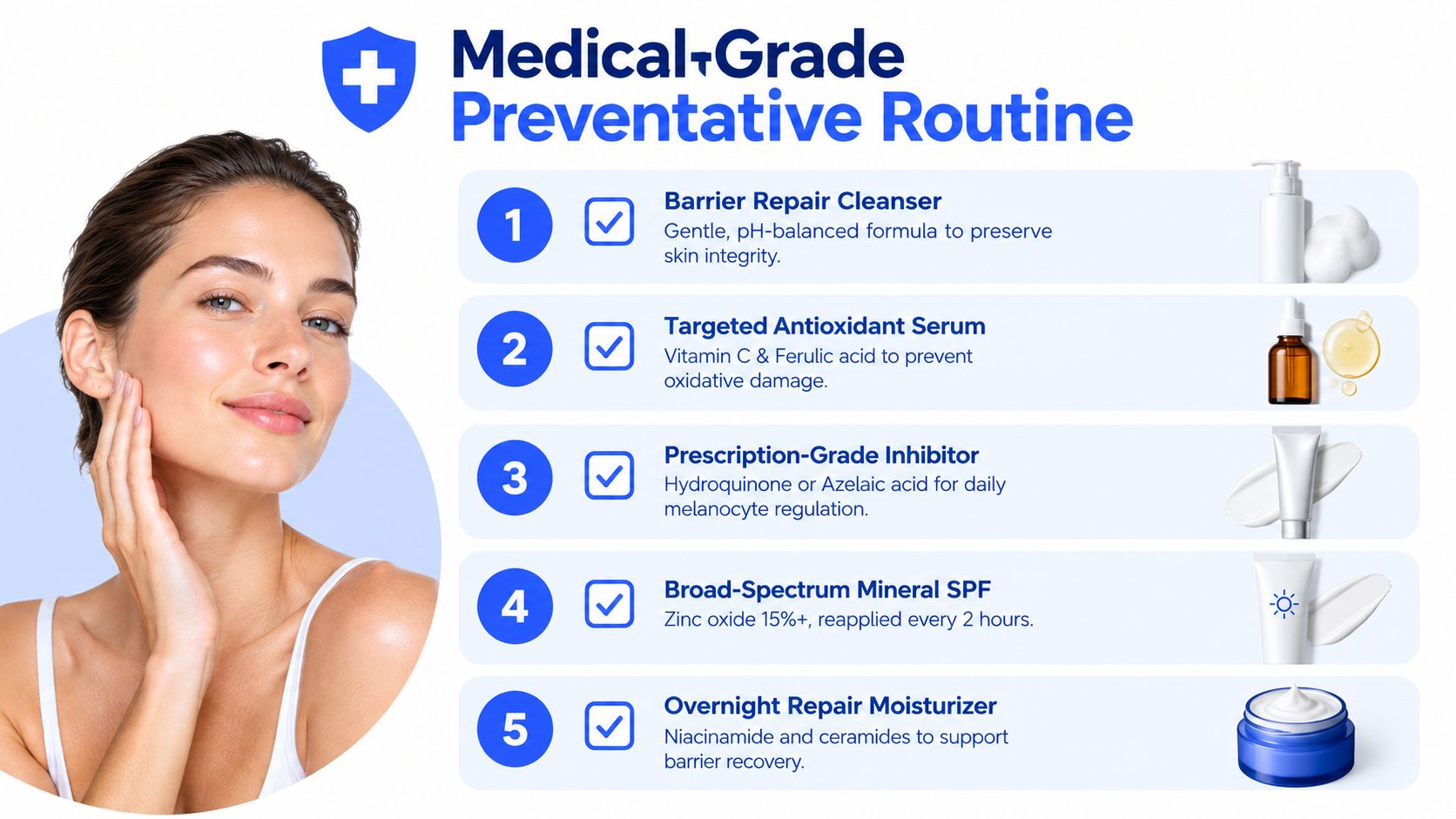
Procedures can address established discoloration, but daily habits determine whether new pigment keeps forming. The foundation is simple: repair the barrier, stop the trigger, protect the area from light, and use targeted actives without creating fresh inflammation.
The 2026 global consensus on melanin hyperpigmentation disorders recommends short-term use of hydroquinone and non-fluorinated steroids such as desonide rather than prolonged use. The global consensus document also emphasizes that certain periorificial and periorbital patterns may be better managed with camouflage, anti-inflammatory care, superficial peels, or low-fluence laser.
A practical daily framework
Morning: Cleanse gently, apply a tolerable antioxidant or pigment-supportive product, moisturize, and apply broad-spectrum sunscreen across the upper lip, corners, chin, and the skin immediately beside the lips. Physical barriers, such as a hat or deliberate shade, add protection when you spend time outdoors.
Evening: Remove sunscreen without scrubbing, use the prescribed or selected pigment-supportive active, and finish with a barrier-focused moisturizer. If the skin stings, flakes, or becomes red, reduce active frequency and address the irritation instead of increasing the dose.
Azelaic acid can be useful when pigmentation overlaps with inflammation or blemish-prone skin. Tranexamic acid, vitamin C, and niacinamide may support different parts of the pigment pathway, but the ideal combination depends on diagnosis and tolerance. The area around the mouth is not a good place to test how many actives the skin can endure.
Maintenance is part of treatment
A provider-curated routine from the ProMD Health medical-grade skincare collection can be used to support in-office care between visits. Products should be selected for the patient's barrier status and diagnosis, rather than purchased solely because a label promises rapid brightening.
Avoid lip licking, abrasive scrubs, unnecessary waxing over irritated skin, and fragranced products that repeatedly touch the surrounding area. If toothpaste or a lip product appears suspicious, a clinician may recommend a structured elimination and reintroduction process. The purpose isn't to strip the routine down forever. It's to find out whether an everyday exposure is maintaining the pigment.
When to Seek Professional Dermatologic Evaluation
Professional evaluation is warranted when the diagnosis is uncertain, the area stays inflamed, or repeated brightening attempts haven't helped. Persistent redness, itching, scale, papules, burning, or cracking suggests that active dermatitis may be driving the discoloration. Treating that skin with stronger bleaching agents before controlling inflammation can make the cycle harder to break.
Seek assessment when pigmentation changes rapidly, develops an unusual border, becomes asymmetric in a new way, bleeds, crusts, or looks unlike the surrounding marks. Drug-induced pigmentation and contact dermatitis also require a medication and exposure review. Rare lesions that need biopsy belong in the differential, which is another reason not to treat every new facial dark spot as a cosmetic concern.
A consultation should answer practical questions:
- What is the dominant cause? Melasma, PIH, contact reaction, constitutional pigment, shadowing, vascular change, or another diagnosis.
- Is the pigment epidermal, dermal, or mixed?
- Is inflammation still active?
- Which treatment carries the lowest risk for the patient's phototype?
- What maintenance plan will reduce recurrence?
ProMD Health provides provider-led aesthetic consultations, skincare, chemical peels, and laser-based options through its approved clinic network, with availability varying by office. Its cash-pay model means patients should confirm the service, location, treatment plan, and payment details directly before scheduling.
ProMD Health can evaluate the cause of your perioral discoloration and build a plan that may combine trigger control, medical-grade skincare, chemical peels, or appropriately selected laser care. Visit ProMD Health to review available services and request a consultation at an approved location.


