
You notice a red patch that never fades, a blue-purple bump that feels soft, or a cluster of fine veins around your nose or legs. It may look like a simple cosmetic concern, but appearance alone can't tell you which treatment will work. Vascular lesion treatment begins with identifying what the lesion is, how it extends, and whether blood flow is slow or fast.
A laser may be appropriate for a superficial capillary lesion, while a deeper venous malformation may respond better to sclerotherapy or a staged combination of treatments. Some lesions need imaging and coordinated evaluation before any procedure is considered.
Core idea: Vascular lesion care is usually iterative. The right plan follows the lesion's biology, not simply its color or the name of a device.
Table of Contents
- Introduction to Vascular Lesions and Why Treatment Choices Matter
- Understanding Vascular Lesions Types Causes and How They Form
- How Vascular Lesions Are Diagnosed and Classified for Treatment Planning
- Non Surgical Vascular Lesion Treatment Options Compared
- Candidacy What to Expect During and After Treatment and Safety Considerations
- Effectiveness Durability Costs and Common Misconceptions About Results
- Next Steps With ProMD Health Locations Services and Telehealth Pathways
Introduction to Vascular Lesions and Why Treatment Choices Matter
A persistent red mark can create several questions at once. Is it a broken blood vessel, a birthmark, a vascular tumor, or a sign of an underlying malformation? Will it disappear on its own, and if treatment is possible, does “removal” mean one appointment or a series?
Those questions matter because vascular lesions aren't one condition. A flat pink patch, a raised red bump, a purple spot on the lip, and a network of blue veins may all look vascular, yet they can differ in vessel type, depth, flow, and natural history. Treating them as interchangeable can lead to disappointing results or unnecessary risk.
The history of pulsed dye laser treatment helped establish a more realistic approach. A review found that about 65% of patients with port-wine stains achieved 50% to 90% lightening, while roughly 15% achieved more than 90% lightening. The same review describes a typical need for 4 to 12 sessions spaced 4 to 6 weeks apart, showing why vascular treatment is often a process rather than a one-and-done procedure (review of pulsed dye laser treatment).
This guide follows the decisions a clinician makes, in plain language:
- Identify the lesion family, such as a vascular tumor or malformation.
- Assess depth and flow, using examination and imaging when needed.
- Match the mechanism to the anatomy, choosing laser, light-based care, sclerotherapy, or a combination.
- Set expectations, including response, recovery, repeat treatment, and possible recurrence.
You don't need to know the diagnosis before seeking advice. You do need an evaluation that treats classification as the first step, because understanding the lesion comes before choosing the procedure.
Understanding Vascular Lesions Types Causes and How They Form
Blood vessels are part of the body's transport system. Arteries carry blood under higher pressure, veins return it at lower pressure, and capillaries form tiny connecting networks near the skin's surface. A vascular lesion develops when vessels grow, widen, cluster, or form abnormally.
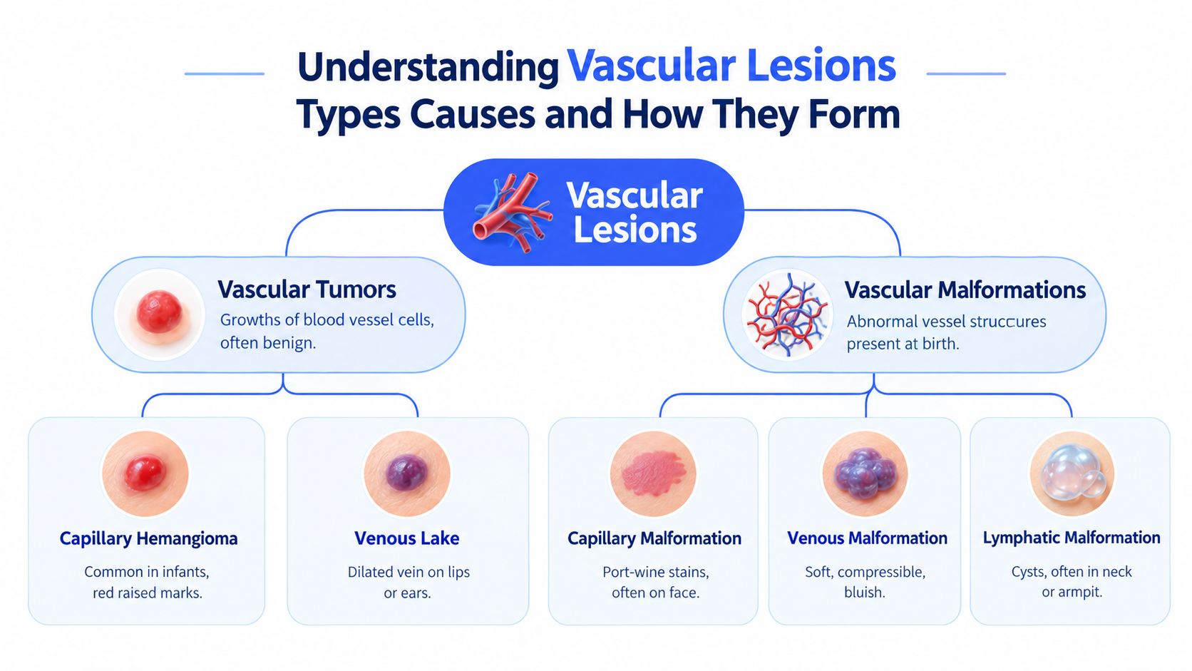
Two broad families
Vascular tumors involve a growth of blood vessel cells. Some are benign and may appear as red, raised marks, such as capillary hemangiomas. A venous lake is another visible vascular concern, typically appearing as a dark blue or purple spot where a vein has become dilated.
Vascular malformations are structural differences in the vessels. They're generally present from birth, even if they become more noticeable later. A capillary malformation, including a port-wine stain, tends to appear as a flat pink, red, or violaceous patch. Venous malformations can feel soft and compressible, while lymphatic malformations contain abnormal lymphatic spaces and may form cyst-like areas.
Why color isn't enough
Think of the skin as a plumbing network with pipes at different depths. A narrow capillary close to the surface may look bright red and respond to a wavelength that targets superficial blood. A larger, deeper vein may look blue or purple because light interacts differently with tissue and blood at depth.
Flow also changes the treatment conversation. A slow-flow venous lesion has a different physical problem from a fast-flow arterial or arteriovenous malformation. The distinction matters because the same visible redness can represent very different vessel architecture.
Causes and triggers vary. Genetics, developmental vessel formation, aging, sun exposure, and other medical factors may contribute, but a visible mark doesn't reveal the entire explanation. The practical question isn't only “What color is it?” It's “What type of vessel created it, and how is that vessel behaving?”
A vascular lesion is better understood as a vessel problem with a visible surface expression, not as a color problem alone.
That shift prevents a common mistake: choosing treatment because a device is advertised for “red spots,” without confirming whether the spot is superficial, deep, slow-flow, or fast-flow.
How Vascular Lesions Are Diagnosed and Classified for Treatment Planning
Diagnosis starts with a conversation and a close examination. A clinician may ask when the mark first appeared, whether it has changed, whether it swells or hurts, and whether symptoms vary with position or activity. Location, texture, compressibility, and color can provide important clues, but complex lesions often need more than visual inspection.
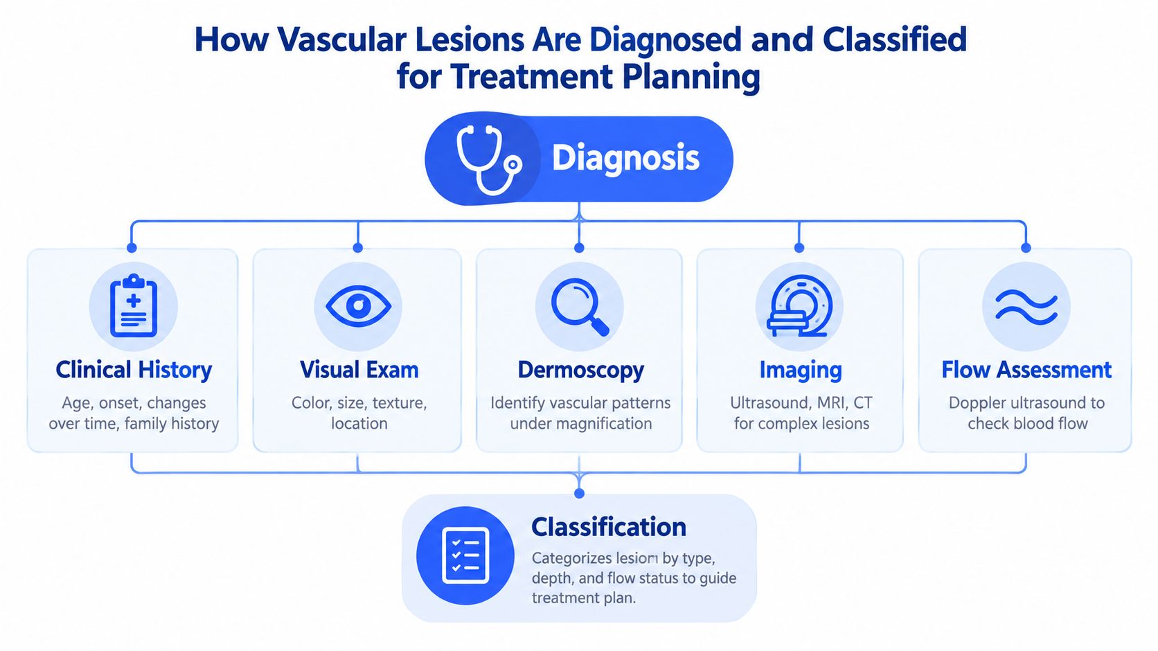
The clinical questions that shape the plan
A typical assessment may include:
- Clinical history, including onset, change over time, symptoms, and relevant family history.
- Visual examination, looking at color, borders, size, texture, and anatomic location.
- Dermoscopy, when magnified vascular patterns can add useful detail.
- Imaging, such as ultrasound, MRI, or CT for lesions that extend deeper or involve complex structures.
- Flow assessment, often using Doppler ultrasound to help distinguish slow-flow from fast-flow anatomy.
These steps answer different questions. Examination describes what the lesion looks like. Imaging shows how far it extends. Flow assessment helps determine whether blood is moving slowly through a venous structure or rapidly through an arterial pathway.
Why classification changes treatment
A superficial lesion with oxyhemoglobin-rich vessels may be a strong laser candidate. A deeper venous malformation may require injection-based treatment because the relevant vessel architecture lies beyond the most effective reach of a superficial laser. Arterial malformations aren't appropriate for vascular laser or IPL treatment, so confirming flow isn't a technical detail. It's a safety boundary.
Complex cases often benefit from coordinated planning. A multidisciplinary vascular anomalies clinic reported that recommendations were needed in 83% of cases, while further diagnostic workup was needed in 43%. In that cohort, recommendations included surgery in 37%, sclerotherapy or embolization in 29%, laser therapy in 12%, and palliative measures such as compression garments or low-dose aspirin in 32% (multidisciplinary vascular anomalies clinic study).
Those figures show why a single-procedure mindset can fail. The correct pathway may involve observation, imaging, laser, sclerotherapy, embolization, surgery, supportive care, or a carefully sequenced combination. Classification protects patients from treating the surface while missing the structure underneath.
Non Surgical Vascular Lesion Treatment Options Compared
Non-surgical options work through different mechanisms, so “laser treatment” isn't a complete treatment plan. The wavelength determines which chromophore absorbs the energy, while spot size, pulse duration, fluence, vessel caliber, skin type, and depth influence how safely that energy reaches the target.
Pulsed dye laser, generally used in the 585 to 595 nm range, targets oxyhemoglobin and has the strongest efficacy and safety evidence for many superficial vascular lesions. Guidelines describe PDL as an option for port-wine stains, hemangiomas, facial telangiectasia, rosacea, spider angioma, pyogenic granuloma, venous lakes, and leg veins (vascular laser guidelines).
KTP at 532 nm is another hemoglobin-targeting option for selected superficial vessels. Intense pulsed light, or IPL, uses a broader band of light rather than one laser wavelength and may be considered for appropriate diffuse redness or superficial vascular concerns. The best choice depends on the lesion and the patient's skin characteristics, not on a general ranking of devices.
Nd:YAG at 1064 nm penetrates more. Reviews describe penetration of about 5 to 6 mm, compared with a maximum penetration depth of about 2 mm for PDL. That depth advantage helps explain why 1064 nm is considered for larger, deeper, or PDL-resistant vessels, including some venous malformations and thick or nodular lesions (Nd:YAG vascular laser review). Another review identifies 532 nm KTP, 585 to 595 nm PDL, and 1064 nm Nd:YAG as established vascular laser families, with IPL included among light-based options (vascular laser wavelength review).
Matching anatomy to modality
| Lesion Type and Depth | Preferred Non Surgical Option | Why It Fits |
|---|---|---|
| Superficial capillary lesion | PDL or selected KTP treatment | Targets hemoglobin near the skin surface |
| Diffuse superficial redness | IPL or an appropriate vascular laser | Can treat broader areas when the pattern and skin profile are suitable |
| Larger or deeper vessel | Long-pulsed Nd:YAG at 1064 nm | Offers greater tissue penetration |
| Low-flow venous malformation | Sclerotherapy, sometimes staged or combined | Treats deeper venous architecture from inside the vessel |
| Low-flow lymphatic malformation | Sclerotherapy or coordinated multimodal care | Addresses abnormal spaces that laser may not reach |
| Fast-flow arterial malformation | Specialist vascular evaluation | Vascular laser and IPL aren't appropriate for arterial malformations |
Sclerotherapy uses an injected sclerosant, such as doxycycline, bleomycin, or sodium tetradecyl sulfate, to injure the vessel lining and trigger thrombosis and fibrosis. This can shrink a lesion and reduce pain, pressure, decompression problems, or visible fullness. Contemporary studies describe these agents as useful across body locations, including intra-orbital lesions, while older clinical series reported success rates above 86% for venous malformations (sclerotherapy review and outcomes).
Electrosurgery may have a role in selected superficial lesions, while topical or light-based adjuncts may support management of the surrounding skin or associated redness. They aren't universal substitutes for lesion-directed therapy.
Practical settings matter too. Expert guidance suggests 10 to 15 mm spot sizes for larger vascular lesions to reduce scatter, with pulse durations around 20 to 30 ms matched to vessel caliber and depth (vascular laser guidelines). In procedural environments, organized imaging and equipment management also matter, which is why facilities may consult resources on durable radiology storage equipment. For small visible vessels, a focused evaluation can help determine whether spider-vein removal is better approached with sclerotherapy, laser, or light-based care.
Candidacy What to Expect During and After Treatment and Safety Considerations
Good candidacy depends on diagnosis, skin characteristics, medical history, medications, lesion depth, and expectations. A clinician should know about pregnancy, active infection, healing problems, a history of abnormal scarring, photosensitivity, recent tanning, and medications that may affect treatment or recovery.
The consultation should answer practical questions:
- What is the working diagnosis? Ask whether the lesion appears to be a tumor, malformation, superficial vessel, or deeper low-flow structure.
- Will imaging be needed? A deeper, painful, enlarging, compressible, or anatomically complex lesion may need further assessment.
- Which endpoint is realistic? The goal may be lightening, volume reduction, symptom relief, or improved contour rather than complete removal.
- What is the sequence? Ask whether laser, sclerotherapy, or staged combination treatment makes sense.
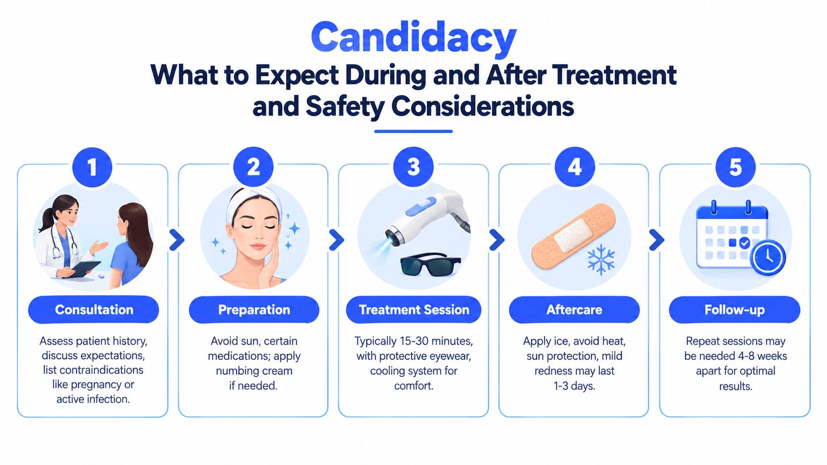
A typical treatment visit
The clinician cleans and examines the treatment area, confirms the plan, and provides protective eyewear. Depending on the modality and location, cooling may be used to improve comfort and protect the epidermis. Patients often describe laser pulses as warmth, tingling, or a brief snapping sensation.
The appointment length depends on the area and lesion complexity. Mild redness, warmth, swelling, tenderness, or bruising can occur afterward. The treated area may look more noticeable before it settles, so immediate appearance isn't a reliable measure of final response.
Recovery and safety
Follow the clinic's specific aftercare instructions. Cool compresses may ease discomfort, while sun protection helps reduce the risk of unwanted pigment changes during healing. Avoiding excessive heat and friction may also be recommended, particularly after treatment of sensitive areas.
Laser parameters are selected to balance target absorption with epidermal protection. Larger spot sizes can reduce scatter for larger vessels, while pulse duration should match vessel caliber and depth. Poorly matched settings can lower efficacy or increase the chance of blistering, pigment alteration, prolonged redness, or scarring.
PDL care often requires repeat treatment. The foundational review cited earlier describes 4 to 12 sessions spaced 4 to 6 weeks apart for port-wine stains (pulsed dye laser review). Deeper lesions may need staged sclerotherapy, imaging-guided care, or another modality rather than increasing laser intensity.
For broader vascular redness and light-based planning, patients may also review how broadband light therapy differs from a focused vascular laser during consultation.
Effectiveness Durability Costs and Common Misconceptions About Results
A lesion can look better after the first treatment without being completely cleared. A superficial vessel may fade, while a deeper malformation may become smaller, less painful, or less congested without disappearing. The appropriate measure of success depends on the lesion's biology and the reason treatment was chosen. Color change alone cannot describe every meaningful improvement.
Evidence from PDL illustrates the range of possible outcomes. About 65% of patients with port-wine stains achieved 50% to 90% lightening, while roughly 15% achieved more than 90% lightening. Patients commonly needed 4 to 12 sessions (pulsed dye laser review). These findings apply to a defined lesion group, not to every red mark or vascular abnormality.
Bleomycin electrosclerotherapy shows why a single cure rate can mislead. An updated systematic review reported complete responses ranging from 65% to 100% in vascular tumors. For vascular malformations, lesion or symptom reduction ranged from 54.9% to 100%, with variation linked to lesion types and study designs (systematic review of bleomycin electrosclerotherapy). The treatment plan must therefore follow classification, flow pattern, depth, and symptoms.
What influences durability
A return of visible vessels does not always mean the same problem has recurred. A treated vessel can reopen, an underlying malformation can remain biologically active, or new vessels can appear with aging, sun exposure, inflammation, or other triggers. Residual disease may also become easier to see after post-treatment swelling subsides.
The important shift in complex vascular care is toward classification and precision. Recent reviews discuss sirolimus, PI3K inhibitors, MEK inhibitors, and genotype-specific treatments, while noting that newer agents still need longer-term safety data (review of targeted vascular malformation therapies). For selected referral patients, these options may affect sequencing with procedures. They do not replace local treatment in every case.
Cost questions without false certainty
Cost varies with lesion size, location, treatment method, clinician time, imaging, and the number of visits. ProMD Health uses a cash-pay model, so patients should confirm whether insurance applies rather than assume reimbursement. Ask about the consultation fee, session charge, expected stages, follow-up policy, and separate pricing for imaging or additional procedures.
Patients comparing IPL with a dedicated vascular laser can review this comparison of IPL and laser treatment to understand why wavelength specificity matters for lesion type. Device choice should follow the target vessel's depth, flow, and absorption characteristics, not the brand name alone.
Three common myths deserve correction:
- “Laser removal always takes one visit.” Many vascular lesions respond gradually and require repeat care.
- “A newer laser solves every vascular problem.” A deep venous malformation may need sclerotherapy or coordinated treatment because vessel architecture, not surface color, drives the problem.
- “Treatment success means the lesion can never return.” Long-term control depends on lesion type, response, and whether residual or new vessels remain.
Next Steps With ProMD Health Locations Services and Telehealth Pathways
A consultation is the appropriate next step when a vascular mark is persistent, changing, symptomatic, raised, compressible, or causing concern. ProMD Health offers laser treatments and a spider-vein pathway that may include laser, light-based treatment, or sclerotherapy, but availability can vary by office and should be verified before booking.
Approved ProMD locations include Annapolis, Arlington, Ashburn, Bel Air, Bethesda, Columbia, Easton, Fort Lauderdale, Hutto, Lafayette, Lewes, Timonium, Tox Bar in Baltimore City, Tysons Corner, Washington, DC, Wellington, and Westminster. The practice uses a cash-pay model, so confirm pricing and payment details directly rather than assuming insurance coverage.
ProMD Health's service themes include lasers, laser hair removal, tattoo removal, injectables, fillers, skincare, spa services, body contouring, hair restoration, weight management, hormone and menopause programs, functional medicine, longevity care, peptide therapy, and ProMD Connect telehealth. Some treatments are office-specific, so ask whether the location you choose provides the vascular service under consideration.
For remote care, ProMD Connect may support consultation and ongoing program management where clinically appropriate. Patients evaluating virtual pathways can also review general telemedicine certification fundamentals. Before scheduling, verify four details: the lesion service, the selected office, the cash-pay estimate, and whether an in-person examination or imaging is needed.
If you're concerned about spider veins, facial vessels, or another visible vascular lesion, visit ProMD Health to review available laser and sclerotherapy pathways. Request a consultation so a provider can assess the lesion's type, depth, and flow before recommending treatment.


